
 Português
  English
  Français
  Русский
  Español
  Deutsch
  العربية
  Italiano
A complete blood count (CBC) is one of the core tests in human hematology. Modern automated CBC systems provide quantitative information on white blood cells, red blood cells, hemoglobin, red-cell indices, platelets, differential results, flags, and graphical outputs, forming an important part of routine hematologic assessment.
Some AI-assisted hematology analyzers add image-based cell morphology information to established automated CBC workflows. The specific testing principle, analyzer configuration, image method, and level of automation vary by model. Where image-based morphology is available, it can provide a complementary visual layer for morphology-related review and make selected cell findings more visible and traceable within laboratory-defined procedures.
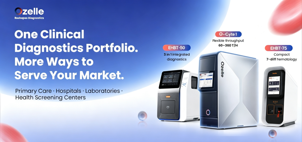
For organizations evaluating compact and automated human hematology systems, Ozelle’s hematology portfolio includes different analyzer architectures, ranging from compact cell morphology analysis and 7-diff hematology to multi-panel testing and modular automated workflows.
Automated CBC as the Quantitative Foundation
Automated CBC is already a mature quantitative testing workflow. Depending on the analyzer architecture, it can provide cell counts, differential information, red-cell indices, platelet-related parameters, flags, and graphical outputs. These outputs support routine hematologic assessment and help laboratories identify samples that may require additional review.
This quantitative foundation should not be reduced to a simple “counts and indices” model. Differential results, flags, histograms, and scattergrams can provide important context for laboratory review. The available information and the review process will vary according to the analyzer, the test menu, and the laboratory’s established procedures.
Cell morphology adds a separate but complementary information layer. It concerns visible cell characteristics such as size, shape, staining appearance, maturation features, and morphology-related abnormalities. When a sample meets laboratory-defined morphology-review criteria, the workflow may include image review, repeat testing, peripheral blood smear preparation, microscopic examination, or other locally defined follow-up procedures.
How an AI Hematology Analyzer Adds a Review Layer
Image-based AI morphology adds visual cell information to automated CBC workflows. Depending on the analyzer design, the system can capture cell images, associate them with morphology-related classifications, and present these findings alongside quantitative hematology data.
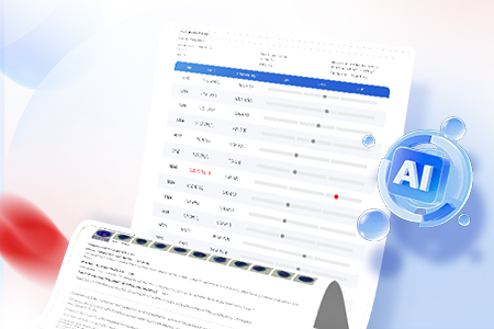
This approach can make selected cell findings more visible during laboratory-defined review. Testing personnel can assess numerical differentials, flags, and graphical outputs together with image-supported morphology-related information as part of the overall result assessment.
The resulting report may include numerical CBC data, morphology-related categories, cell images, and graphical outputs. Together, these elements can support a more traceable review process, particularly when results trigger laboratory review criteria or contain morphology-related flags.
Where available, AI-assisted morphology can make morphology-related visual information accessible within the same reporting and review process as CBC data. Qualified laboratory interpretation and clinical correlation remain part of routine hematology practice.
How Image-Based Morphology Fits into CBC Workflows
Automated CBC, Flags, and Review Rules
Automated CBC results can include quantitative cell counts, differential data, red-cell and platelet indices, flags, and graphical outputs. Laboratories can use locally defined review rules to determine which results require additional examination.
Review criteria may be triggered by instrument flags, unusual cell distributions, unexpected count patterns, or other laboratory-defined parameters. The follow-up process varies by institution and may include result verification, repeat analysis, image review, or peripheral blood smear examination.
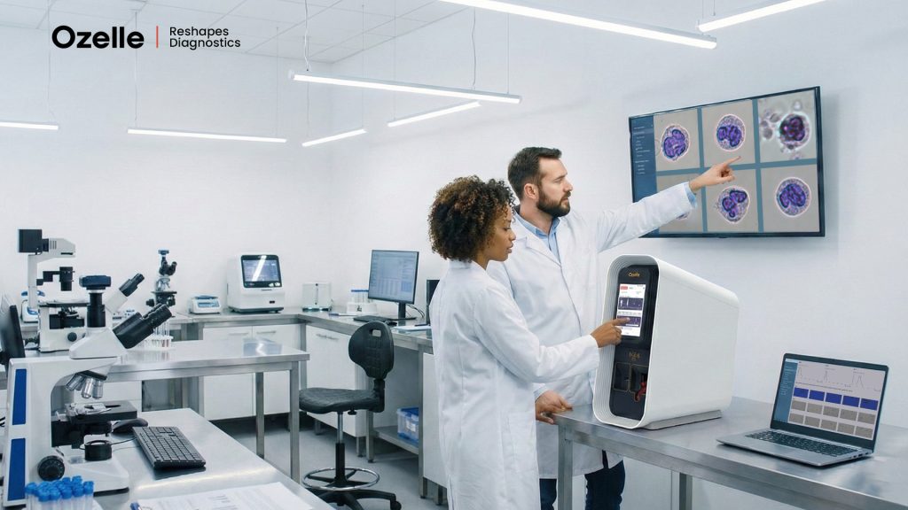
Image-Based Morphology Review
Image-based morphology adds visual cell information to this review process. Cell images and associated classifications can help testing personnel examine morphology-related findings in a more visible and traceable format.
Not every sample requires additional manual review of the available cell images. Instead, image-based morphology can be incorporated into selected workflows where laboratories need additional information beyond quantitative CBC results, flags, and graphical outputs. This allows the laboratory to apply image-supported morphology according to its own review criteria and service scope.
Qualified Interpretation and Clinical Correlation
Hematology results are reviewed according to laboratory procedures and interpreted alongside relevant clinical information. Testing personnel evaluate analyzer output according to local policies, while clinical teams consider laboratory findings alongside patient presentation, medical history, and other relevant investigations.
This structure places AI-assisted morphology within a defined review process rather than treating it as a separate diagnostic pathway. Image evidence, numerical results, laboratory procedures, and clinical correlation each contribute different information to the overall assessment.
Hematology Analyzer Architectures for Different Workflows
Image-based morphology and automated hematology do not require a single analyzer format. Laboratories differ in workload, available space, required test menus, sample-handling processes, connectivity needs, and quality-management procedures.
Ozelle’s human hematology portfolio includes analyzers with different testing principles, hematology configurations, integrated test capabilities, and automation formats. These systems are designed for different laboratory workflows, ranging from compact cell morphology analysis and 7-diff hematology to multi-panel testing and modular automated hematology.
EHBT-25 for Compact Morphology-Oriented Testing
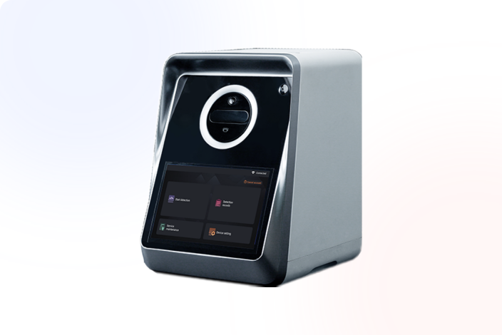
O EHBT-25 is a compact 3-diff cell morphology analyzer based on cell morphology imaging technology and photoelectric colorimetry. It captures and displays actual WBC, RBC, and platelet images, providing morphology-related visual information alongside routine CBC results.
EHBT-25 uses capillary or venous whole blood and has a documented throughput of eight samples per hour. Its specified configuration includes a dry-type QC card and manual calibration. This architecture may suit primary and community healthcare settings that need a compact morphology-oriented analyzer and a defined workflow for handling results requiring further laboratory review.
EHBT-50 for Compact Multi-Panel Testing
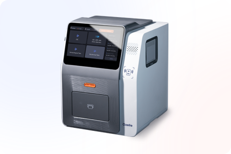
O EHBT-50 is a compact multi-functional analyzer that combines 7-diff CBM, immunoassay, and biochemistry testing. Users can configure single, dual, or triple test combinations in one batch, allowing the testing menu to be matched with the actual needs of the facility.
Its hematology configuration includes standard CBC data and extended categories including NST, NSG, NSH, ALY, PAg (platelet aggregate-related parameter), and reticulocytes. The system also includes derived CBC indices such as NLR and PLR. These indices should be understood as calculated hematology parameters derived from CBC data, rather than as morphology findings.
EHBT-50 supports capillary or venous whole blood for hematology testing, as well as serum and plasma for relevant immunoassay and biochemistry panels. Its documented configuration includes dry-type QC card and liquid QC options, auto calibration, LIS connectivity, and a throughput of eight samples per hour. These features should be evaluated according to the laboratory’s own workflow, quality-management process, and expected sample volume.
EHBT-75 for 7-Diff Morphology Workflows
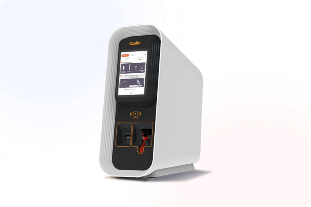
O EHBT-75 is an AI-powered 7-diff auto hematology analyzer. Its workflow combines liquid-based staining, image-based detection, high-resolution imaging, and AI-assisted cell classification.
The analyzer uses capillary or venous whole blood and reports standard CBC parameters alongside categories including NST, NSG, NSH, ALY, platelet aggregates, and reticulocytes. Its documented throughput is 7–8 samples per hour, supporting compact human hematology workflows where image-based cell morphology is incorporated into routine CBC testing.
O-Cyte 1: High-Throughput AI Hematology with Modular Expansion
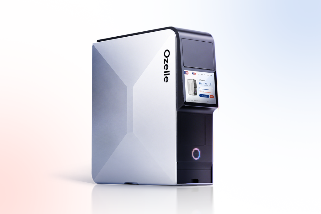
O-Cyte 1 is an automated hematology analyzer combining 7-diff CBC with AI-assisted morphology in one workflow. Its modular architecture supports standalone operation or cascaded expansion, with a documented throughput of up to 60 tests per hour as a standalone unit and up to 360 tests per hour through cascaded expansion.
The analyzer includes a 25-position batch-loading configuration, automatic barcode identification, automated sample preparation, image-based morphology analysis, AI-assisted classification. It also supports LIS/HIS, USB, RJ45, Wi-Fi, and Bluetooth connectivity.
For laboratories with larger CBC workloads, this architecture can make morphology-related image information available within an automated workflow that includes batch handling, analyzer connectivity, and scalable capacity. O-Cyte 1 also lists internal morphology liquid QC support, reflecting a quality-control configuration specific to that model.
Model Selection Criteria
When comparing an AI hematology analyzer or another morphology-oriented hematology system, laboratories should base model selection on actual workflow requirements rather than technology labels alone.
| Evaluation area | Questions to assess | Por que é importante |
| Hematology scope | Does the analyzer provide the required CBC, differential, morphology, or multi-panel workflow? | The analyzer should align with the facility’s testing service scope |
| Morphology information | Does the system provide cell images, morphology-related categories, or image-supported classifications? | Determines how morphology-related information enters review procedures |
| Workload and analyzer format | Is a compact, single-analyzer, or modular higher-throughput configuration needed? | Connects capacity planning with routine and peak sample volumes. |
| Sample handling | Which sample types and loading approaches are validated for the selected model? | Prevents generalizing venous or capillary workflows across all systems |
| Quality management | What QC and calibration methods are specified for the individual model? | QC and calibration are model-specific rather than portfolio-wide features |
| Conectividade | Does the analyzer support LIS, HIS, or other required reporting interfaces? | Helps integrate the analyzer into laboratory reporting workflows |
| Operational fit | What are the training, consumable, space, maintenance, and service requirements? | Supports sustainable day-to-day implementation |
The appropriate system depends on how the laboratory intends to use morphology-related information within its CBC workflow. A compact 3-diff morphology analyzer, compact 7-diff analyzer, multi-functional system, and scalable automated platform each serve different operational requirements.
Conclusão
Modern automated CBC already provides a strong quantitative foundation for routine hematology testing. In selected workflows, an AI hematology analyzer can add a complementary image-based morphology layer that makes cell findings more visible, traceable, and easier to incorporate into laboratory-defined review procedures.
The appropriate analyzer architecture depends on workload, available space, required test menu, sample-handling process, quality-management procedures, and connectivity needs. Laboratories can evaluate compact morphology analysis, integrated multi-panel testing, 7-diff image-based hematology, and modular automation according to how each approach fits their established CBC workflow.
For laboratories assessing how AI-assisted image-based morphology may fit into their CBC review process, Ozelle’s human hematology portfolio provides options across different workflow architectures.