
 Português
  English
  Français
  Русский
  Español
  Deutsch
  العربية
  Italiano
AI-assisted morphology analysis combines image-based cell evaluation, automated image acquisition, algorithm-supported recognition, and structured result presentation in hematology workflows. It brings cell-level visual information into automated hematology testing alongside CBC parameters, leukocyte differentials, flags, histograms, and other analyzer outputs.
In integrated image-based systems, this can bring cell-level visual information together with CBC parameters, leukocyte differentials, flags, histograms, and other analyzer outputs within the same review workflow.
This approach is relevant to laboratories seeking more structured access to morphology-related information in routine CBC workflows.

Morphology Analysis in Hematology
Blood-cell morphology analysis examines visible cellular characteristics that complement quantitative hematology results. Depending on the analytical method and cell type, the assessment may include cell size, shape, nuclear structure, cytoplasmic appearance, granularity, maturity-related characteristics, and the distribution of cell populations within a sample.
Leukocyte morphology can include differences in nuclear segmentation, chromatin pattern, cytoplasmic granulation, and the appearance of cells at different stages of maturation. Red-cell review can consider variation in size and shape, while platelet-related review can examine platelet appearance, distribution, and aggregation patterns when those features are represented in the analytical output.
Depending on the validated capabilities of the system, morphology analysis may organize cell images into candidate categories and highlight selected cell populations or morphology-related findings for closer review. This can be useful when a laboratory needs to evaluate whether a numerical pattern is accompanied by visible cellular features that add context to the hematology result.
When considered alongside CBC parameters, leukocyte differentials, histograms, flags, and other laboratory information, morphology analysis can add visible cell-level context to laboratory review and subsequent clinical assessment. This creates a broader information set for reviewing hematology results within routine laboratory operations.
Practical Applications of AI-Assisted Morphology Analysis
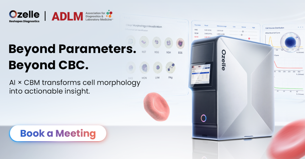
How AI-assisted morphology enters the workflow depends on the system architecture. In downstream digital morphology workflows, CBC findings or laboratory review rules may determine which samples proceed to imaging. In integrated image-based hematology systems, cell images and AI-assisted classifications may already be generated as part of the primary analytical workflow, while laboratory rules determine which results require additional professional review. AI-assisted morphology analysis can support laboratories that need structured access to cell-level images during hematology result review. It is particularly relevant when CBC patterns, differential results, analyzer flags, or laboratory-defined action criteria indicate that a sample requires closer assessment.
In routine hematology operations, image-based results can support the review of leukocyte classifications, cellular maturity patterns, red-cell appearance, platelet-related findings, and other morphology-related information. Depending on their validated scope, AI-supported systems may organize cell images into candidate categories and present selected morphology-related findings for review. AI-supported systems can organize candidate cell categories and highlight selected findings, allowing laboratory staff to view cellular images with quantitative outputs.
This arrangement can be useful where laboratories need to manage large numbers of routine CBC results while maintaining a consistent process for selected morphology-related findings. Rather than reviewing images as an isolated data source, laboratory staff can consider cell-level information within the same result environment as differential values, flags, and graphical outputs.
For laboratory managers, this approach can be evaluated as a workflow resource as well as an analytical capability. The operational question is how image-based cell information fits existing review pathways, staffing structures, documentation practices, and result-reporting requirements.
Requirements for Reliable Review
AI-assisted morphology analysis involves connected stages of sample processing, image generation, classification, result presentation, and laboratory review. The following factors shape how image-based morphology information is incorporated into an automated hematology workflow.
| Requisito | Why it matters | Practical laboratory consideration |
| Sample quality and handling | Sample preparation affects the condition of cells available for image-based analysis | Establish suitable sample acceptance, handling, and timing procedures |
| Cell imaging conditions | Focus, contrast, cellular separation, and overlap affect visible cell-level detail | Assess image consistency and the availability of individual-cell images |
| Image-based classification | Algorithms organize cell images into candidate groups and highlight selected patterns | Define how classifications are reviewed, documented, and escalated |
| Result traceability | Cell images can be reviewed with the classifications or findings shown in the result output | Confirm that visual evidence is accessible with parameters and graphical outputs |
| Review criteria | Morphology-related outputs need defined actions and reporting pathways | Align analyzer outputs with local CBC review criteria and laboratory policies |
| Quality control and training | Stable operation and consistent result review depend on suitable oversight | Define QC procedures, staff responsibilities, and competency assessment |
| Connectivity and records | Image-based results need to integrate with laboratory information processes | Assess barcode workflows, data traceability, storage, and LIS connectivity |
| Verification / validation | Classification and morphology outputs must be understood within the system’s validated performance | Define local verification or validation requirements before routine reporting |
ICSH guidance on digital blood-smear morphology emphasizes the need to consider pre-analytical, analytical, and post-analytical factors together, including staining, image acquisition and display, software, classification performance, and result handling. These recommendations primarily address digital blood-smear morphology systems; integrated image-based CBC analyzers may use different analytical architectures and should be evaluated according to their own intended use, validated performance, and laboratory procedures.
AI-Assisted Morphology Supports Review, Not Autonomous Diagnosis
ICSH guidance on digital blood-smear morphology emphasizes the need to consider pre-analytical, analytical, and post-analytical factors together, including staining, image acquisition and display, software, classification performance, and result handling. These recommendations primarily address digital blood-smear morphology systems; integrated image-based CBC analyzers may use different analytical architectures and should be evaluated according to their own intended use, validated performance, and laboratory procedures.
AI-Assisted Morphology Analysis With O-Cyte 1
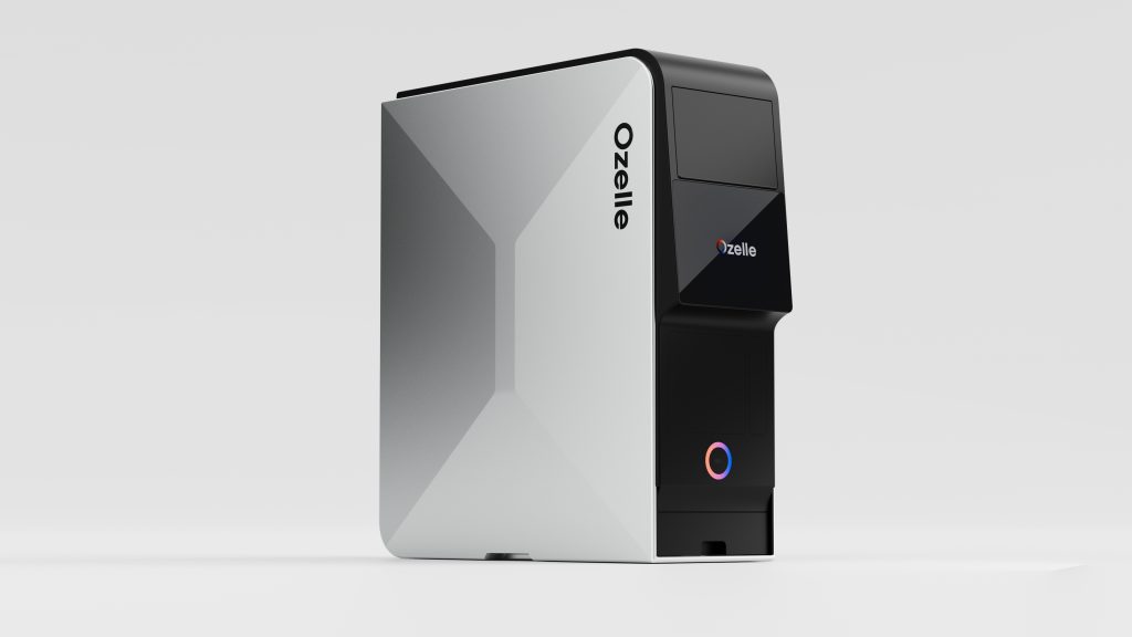
O Analisador hematológico automatizado O-Cyte 1 applies Ozelle’s AI × Complete Blood Morphology (CBM) approach by combining 7-diff CBC with AI-assisted morphology in one workflow. It integrates cell imaging, AI-assisted blood-cell recognition and classification, and image-backed morphology information with routine hematology results.
The analyzer presents cell images, AI classifications, histograms, highlighted findings, and morphology-related outputs in one result display. The system links quantitative results and morphology-related findings to corresponding cell images, allowing reviewers to examine image-based evidence alongside CBC and differential information.
O-Cyte 1 reports CBC and differential parameters including NST, NSG, NSH, ALY, NLR, and PLR, with RET# and RET% available as optional parameters. For laboratories evaluating analyzer outputs, the relevant consideration is the relationship between morphology information and the rest of the hematology result. A display that combines cell images, highlighted findings, parameters, and graphical information can support a more organized review of the available evidence.
O-Cyte 1 in Laboratory Settings
O-Cyte 1 is designed for automated hematology workflows that combine routine CBC testing with image-based morphology information. It accepts whole blood, capillary blood, and prediluted samples, while its Auto Loader Mode supports 25 samples across five racks; automatic barcode identification, sample mixing, closed-tube piercing, and STAT handling are incorporated into the sample-processing sequence.
This configuration is relevant for laboratories that need to process structured batch processing of routine CBC samples while accommodating priority testing within the same workflow. The analyzer supports up to 60 tests per hour in standalone operation and up to 360 tests per hour in cascaded configurations, providing capacity options for routine volume, concentrated demand periods, and STAT sample handling.
In practice, the suitability of this workflow depends on how sample receipt, CBC review, image-based morphology review, and result reporting are organized within the laboratory. For distributors and local partners, implementation can therefore be assessed against the site’s expected workload, laboratory information workflow, training arrangements, and service model.
Conclusão
AI-assisted morphology analysis integrates cell images, algorithm-supported recognition, candidate classification, and structured result presentation into automated hematology workflows. It adds morphology-related evidence to the quantitative and graphical information used in laboratory review and broader clinical assessment.
For laboratories planning an image-based hematology workflow, key considerations include image quality, validated classification performance, clear image–result linkage, alignment with local review criteria, and integration with reporting processes. These factors help determine how image-based morphology information is incorporated into daily hematology operations.
If you are assessing whether O-Cyte 1 is suitable for your laboratory workflow, contactar Ozelle or email info@ozellepoct.com. The Ozelle team is available to answer your questions about O-Cyte 1 and its application in your laboratory workflow.