

In most hospitals and clinics, the hematology auto analyzer is often one of the first analytical instruments used after sample collection, providing essential information to support clinical assessment of infection, anemia, bleeding risk, and systemic conditions. Traditional systems have become very fast and reliable at counting cells, yet they still fall short when clinicians need nuanced morphology and early pattern recognition rather than just numbers.
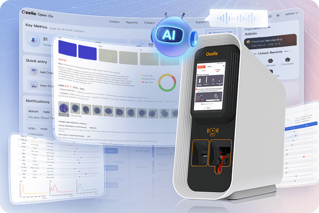
Artificial intelligence is closing this gap. AI-powered hematology auto analyzers combine high‑resolution imaging with deep learning models trained on millions of blood cell images, turning each CBC into a morphology‑aware, interpretation‑ready report. Ozelle’s EHBT‑75 is a representative example of this new generation: a compact 7‑differential hematology auto analyzer that uses AI to provide deep cell morphology from a single drop of blood.

From Conventional CBC to AI-Powered Hematology Auto Analyzer
A conventional hematology auto analyzer uses electrical impedance and optical scatter to count and size cells. This works well for parameters such as WBC, RBC, HGB, PLT and basic differentials. However, the analyzer does not truly “see” cell shapes, nuclear segmentation, or cytoplasmic changes. When results are abnormal, it generates flags and the lab falls back to manual smears, staining, and microscopic review.
In busy settings, this dependency on manual morphology causes delays and inconsistency. Early blasts in leukemia, subtle left shifts in bacterial infections, or hypersegmented neutrophils in megaloblastic anemia may be overlooked or recognized late if the smear is not reviewed promptly or if expert morphologists are not available.
An AI-enabled hematology auto analyzer changes this workflow. Instead of relying only on electrical signals and scatter plots, the instrument captures digital images of cells, feeds them into a deep learning model, and receives a classification and morphology interpretation in return. On Ozelle’s analyzer, this is called Complete Blood Morphology (CBM). The system uses a high‑precision optical path, liquid‑based cytology staining, and convolutional neural networks trained on over 40 million clinical samples. The AI engine distinguishes standard white cell populations and their maturation stages, recognizes atypical lymphocytes, evaluates RBC shapes, and detects platelet aggregates with a level of consistency that is difficult to achieve manually in routine practice.
This shift moves the hematology auto analyzer from a pure counting device towards an intelligent observer that can provide both quantitative and qualitative insight in a single report.
EHBT‑75: AI Hematology Auto Analyzer Centered on Morphology
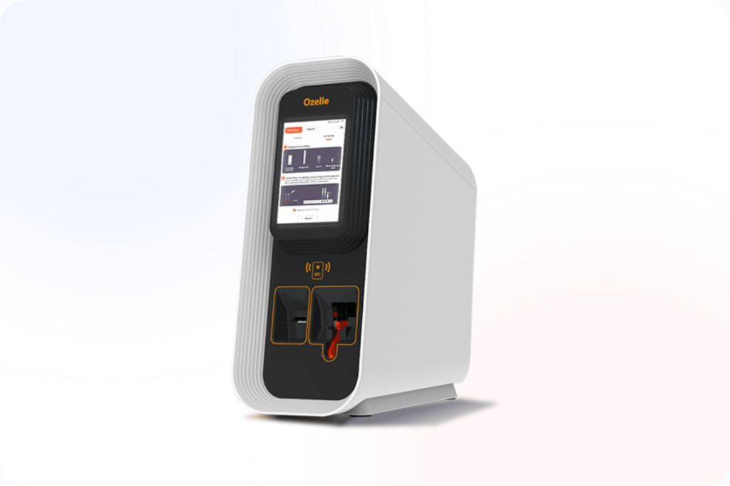
The Ozelle EHBT‑75 hematology auto analyzer is designed with a single purpose: make advanced morphology analysis more accessible within routine CBC workflows. Instead of relying only on electrical signals and scatter plots, this 7‑differential analyzer uses high‑resolution imaging and deep learning so its AI engine “looks at” real cell images before generating results.
In practice, EHBT‑75 behaves like an all‑in‑one station for one blood sample. It accepts a micro‑volume of whole blood, processes it in a closed, single‑use cartridge, performs liquid‑based staining, and captures detailed images of white cells, red cells, and platelets. Those images are analyzed by Ozelle’s CBM model, trained on tens of millions of clinical samples, and converted into a morphology‑aware report that combines CBC, 7‑part differential, key morphology parameters, and inflammation ratios such as NLR and PLR.
Because this entire chain is automated, most samples no longer need a smear before clinicians can see meaningful morphology patterns. The compact 415 mm × 203 mm × 483 mm bench‑top footprint makes it easy to place EHBT‑75 in emergency departments, oncology units, and satellite labs that need fast, morphology‑rich results without building a dedicated morphology lab.
EHBT‑75 Workflow: From Micro-Sample to AI CBC Report
- The operator loads 30–60 μL of venous or capillary whole blood into a single‑use, room‑temperature cartridge and inserts it into the analyzer.
- EHBT‑75 automatically performs mixing and liquid‑based staining in the closed cartridge, then acquires high‑resolution and Z‑stack images of WBC, RBC, and PLT.
- A deep learning engine classifies cells and generates a CBC + 7‑diff + morphology‑rich report—including parameters such as NST, NSG, NSH, ALY, PAg, RET and ratios like NLR and PLR—typically in about six minutes.
Technology Comparison: Traditional vs AI Hematology Auto Analyzer
| Aspetto | Microscopia manuale | Conventional Analyzer | AI Hematology Auto Analyzer (EHBT‑75) |
| Core method | Expert visual review of stained smear | Impedance and optical scatter counting | Digital imaging plus deep learning morphology |
| Output | Detailed but subjective morphology | Fast counts and basic differentials | CBC + 7‑diff + advanced morphology parameters |
| Volume del campione | Several hundred µL | 50–200 µL typical | 30–60 µL venous or capillary blood |
| Tempi di elaborazione | 45–60+ minutes including smear | 10–20 minutes plus smear if flagged | About 6 minutes including morphology |
| Coerenza | Depends on reader and workload | Good for counts, limited for morphology | High reproducibility across time and sites |
| Workload | Labor‑intensive, requires specialists | Moderate; smear work still high | Smear workload significantly reduced |
These characteristics reflect data and capabilities described in Ozelle’s hematology and EHBT‑75 materials.
How AI in EHBT‑75 Delivers Real Clinical Value
Enhanced Infection Assessment
Traditional analyzers may flag a “left shift” but require manual smear review to confirm. EHBT‑75’s AI‑driven cell morphology quantifies immature granulocyte subsets (NST#, NSG#) and provides ratios like NLR directly in the automated report. This offers immediate, objective evidence of neutrophil mobilization, helping clinicians suspect serious bacterial infection or systemic inflammation sooner, without waiting for manual microscopy.
Sensitive Detection of Cellular Abnormalities
Early blood disorders can manifest with subtle morphological changes. EHBT‑75’s high‑resolution imaging and AI analysis are designed to identify atypical cells—such as blasts or dysplastic forms—with high sensitivity. By highlighting these abnormalities and providing visual cell images, the system prompts clinicians to consider timely hematology consultation and further specialized testing for concerning cases, potentially accelerating the diagnostic pathway.
Informed Anemia & RBC Disorder Workup
Beyond standard indices like HGB and MCV, EHBT‑75 integrates reticulocyte count (RET%) and AI‑based RBC morphology analysis. It can automatically flag the presence of abnormal shapes, such as schistocytes or teardrop cells, which are key indicators of hemolysis or marrow pathology. This enriched contextual information aids in differentiating between causes of anemia (e.g., production vs. destruction) directly from the initial CBC, guiding more targeted follow‑up.
Workflow Efficiency and Near-Patient Placement
From a workflow perspective, the combination of micro‑volume sampling, six‑minute turnaround, and room‑temperature cartridges means EHBT‑75 can be located where time matters most, such as emergency departments or short‑stay units. The same instrument that provides deeper morphology may help streamline workflows in emergency settings and relieve pressure on central labs by delivering decision‑ready results at the point of care. With USB, Ethernet, Wi‑Fi, and Bluetooth connectivity, EHBT‑75 connects to LIS/HIS and Ozelle’s IoT analyzers, giving facilities both richer clinical information and a smoother, more predictable diagnostic process across their network.
EHBT‑75 in Ozelle’s AI Hematology Ecosystem
EHBT-75 is part of a broader hematology portfolio rather than a standalone system. It operates alongside multi-functional analyzers and 3-part systems built on the same AI × CBM framework, enabling more consistent morphology-related outputs and interpretation across different clinical settings.
Within this portfolio, EHBT-75 is positioned for scenarios where detailed hematology analysis is required—such as emergency departments, oncology units, and acute care settings. Other Ozelle systems can be deployed in primary care or high-throughput screening environments, allowing institutions to align testing capability with clinical demand and workload.
For detailed specifications and clinical applications, refer to the EHBT-75 product page on Ozelle’s official website: https://ozellemed.com/en/ehbt-75/
FAQs: AI Hematology Auto Analyzer and EHBT‑75
What kind of training do staff need to safely use EHBT‑75 in ED or satellite labs?
Because EHBT‑75 automates loading, staining, imaging, and AI analysis inside a closed cartridge, day‑to‑day operation mainly involves correct sampling, cartridge insertion, and basic QC checks rather than manual smear or complex method steps. Ozelle typically recommends a short on‑site or remote training session focusing on sample collection, result interpretation (including morphology parameters and flags), and integration with the hospital’s LIS, which is generally easier to adopt than training new staff to read blood films under a microscope.
How are AI updates and algorithm validation handled over the lifetime of the analyzer?
EHBT‑75’s morphology engine is based on a continuously improving CBM model, but updates are not pushed blindly. Ozelle validates new algorithm versions on large, multi‑center datasets before release and typically delivers them as versioned firmware or software updates, accompanied by documentation so labs can understand what changed and, if needed, perform local correlation or verification according to their internal quality procedures.
What quality control (QC) and regulatory practices support AI-based morphology results?
From a regulatory standpoint, EHBT‑75 is CE‑marked and developed under ISO‑aligned quality systems, so its core CBC measurements and workflows follow recognized IVD standards. For AI morphology, Ozelle recommends combining standard hematology QC materials with periodic cross‑checks against reference methods or expert reviews, especially when a lab first adopts the system, so the AI output is integrated into existing QC and accreditation frameworks rather than treated as a “black box.”