

Introduction: Why automated animal hematology analyzers matter in veterinary care
In contemporary veterinary medicine, laboratory data have become essential for diagnosing and managing infectious, inflammatory, hematologic, and systemic diseases in animal patients.
A complete blood count supports clinicians in assessing leukocyte responses, anemia patterns, and platelet status, which together provide an early signal of acute infection, chronic inflammation, or bone marrow stress in dogs and cats.
As case volumes rise and pet owners expect faster, evidence-based decisions, the shift from manual microscopy to the automated animal hematology analyzer has become a structural trend in veterinary diagnostics.
At the same time, the veterinary diagnostics market is expanding, increasing demand for automated systems that deliver standardized, connected, and interpretable results across animal hospitals and diagnostic laboratories.
From manual microscopy to early automated veterinary hematology
Historically, veterinary hematology relied on manual blood smear preparation and microscopic review, requiring trained personnel to identify cell types and assess morphology visually.
This approach provided rich qualitative insight but was time-consuming, highly dependent on operator skill, and difficult to standardize across different veterinary settings.
As practice sizes and sample volumes grew, inconsistency in manual counts and morphology interpretation became an operational constraint in many animal hospitals and laboratories.
The introduction of impedance-based and flow cytometry-based analyzers helped address some of these challenges.
Impedance methods estimate cell number and volume from electrical resistance changes, while flow cytometry uses hydrodynamic focusing and laser-based light scattering to distinguish cell populations.
These technologies improved throughput and quantitative precision for many parameters in veterinary CBC testing.
However, they rely largely on indirect physical signals rather than direct visualization of cells, so subtle morphological abnormalities, immature cells, or dysplastic changes often still require manual microscopic review.
This gap motivated the development of image-based and AI-enabled automated animal hematology analyzers tailored to veterinary needs.
The shift to image-based AI for animal blood cell morphology
From signal-based counting to image-based analysis
Traditional automated hematology in veterinary medicine mainly relied on impedance and flow cytometry, which infer cell types from electrical resistance and light-scatter signals.
These methods improved throughput but still could not directly visualize canine and feline blood cells, so subtle morphological abnormalities often required manual microscopic review.
This limitation created a need for technologies that could combine automation with morphology-level insight in the automated animal hematology analyzer.
Core concepts of Complete Blood Morphology (CBM) in veterinary analyzers
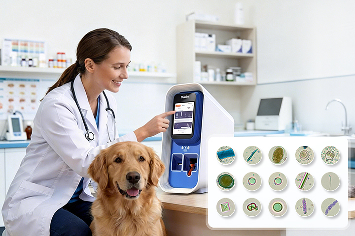
Complete Blood Morphology (CBM) introduces a different approach by capturing high-resolution images of blood cells and using convolutional neural networks to classify them.
Veterinary analyzers such as the veterinary multi-functional hematology analyzer EHVT-50 integrate standardized wet-staining, a dedicated optical path, and AI models trained on large datasets of canine and feline samples.
This allows the system to differentiate leukocyte subtypes, red cells, and platelets while preserving the visual evidence behind each classification.
Optical innovations supporting AI morphology
To support CBM, modern analyzers combine customized high-resolution lenses with high-speed image acquisition and multispectral illumination.
Z-stack scanning acquires images at multiple focal planes, enabling three-dimensional reconstruction of cells and stable focus even in thick or uneven smears.
These optical innovations provide the detailed, consistent input that AI algorithms require to recognize immature neutrophils, fragmented erythrocytes, and other subtle abnormalities in animal blood.
Impact on veterinary diagnostic workflows
By shifting from purely signal-based methods to image-based AI morphology, the automated animal hematology analyzer can reduce the number of samples requiring manual microscopic review.
Routine canine and feline CBCs, including many mild to moderate inflammatory or anemic presentations, can be processed with automated differential counts and morphology flags, while only the most complex cases are escalated for manual review.
This rebalances the workload in veterinary laboratories and supports more consistent interpretation across different sites.
Architecture of a modern veterinary automated animal hematology analyzer
Sample handling and cartridge-based workflow
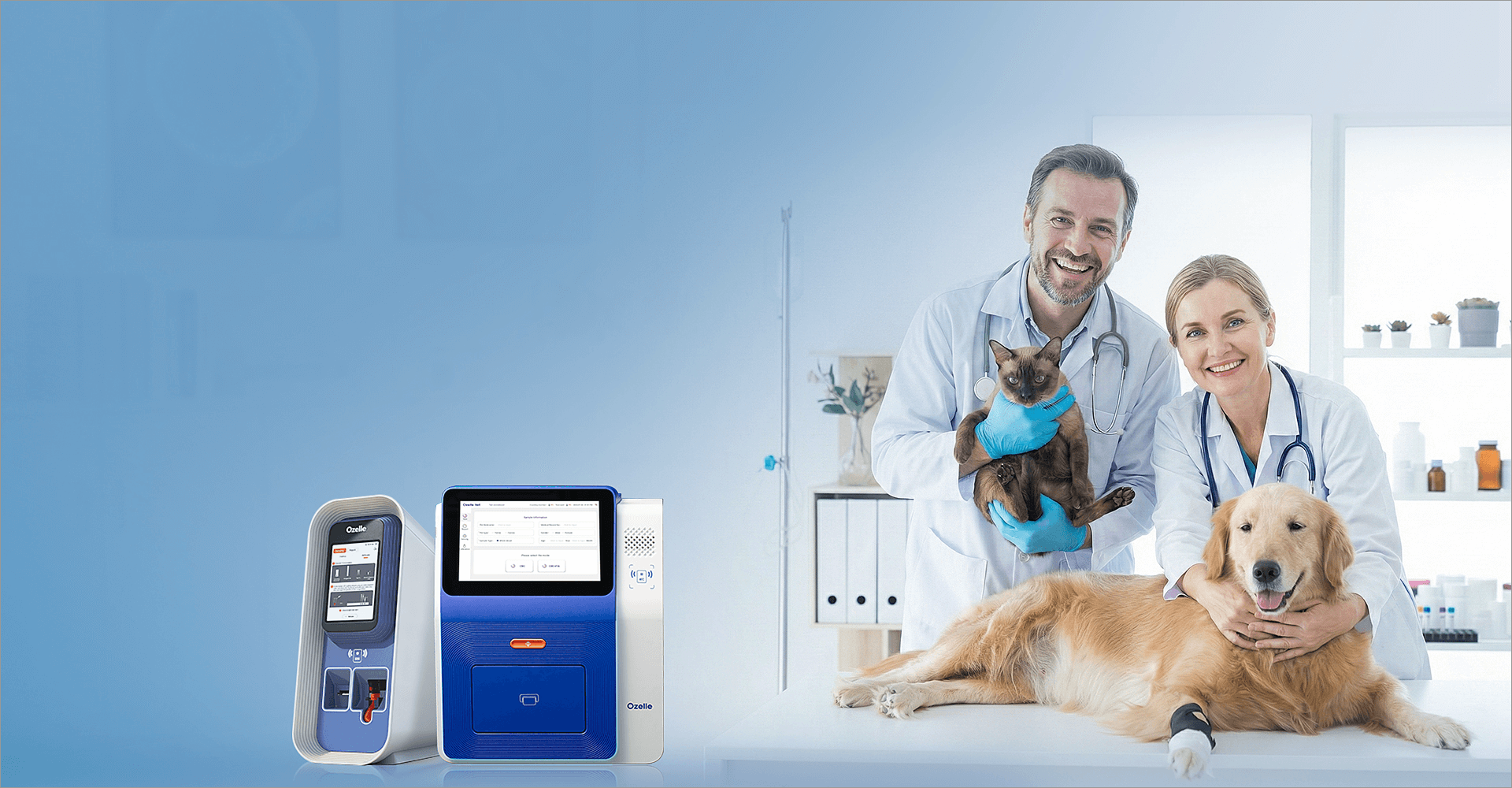
A modern veterinary automated animal hematology analyzer starts with automated sample handling, drawing whole blood from EDTA tubes or capillary collections.
Instruments such as EHVT-50 dispense the sample into single-use test kits, mix it with reagents, and perform staining without manual pre-treatment.
Sealed cartridges and internal waste storage limit operator exposure and reduce routine maintenance needs in veterinary clinics.
Imaging and illumination system
The imaging subsystem combines a high-resolution optical lens with controlled illumination and high-speed image acquisition.
Multispectral light sources enhance contrast for different cell types, while scanning mechanisms ensure that the entire field is captured at the appropriate focus.
This design provides the consistent image quality required for AI-based morphology analysis.
AI engine and morphology classification
Once images are captured, an embedded AI engine performs cell detection, segmentation, and classification across leukocytes, erythrocytes, and platelets.
The model also flags atypical morphology and abnormal distributions that may warrant closer review by veterinary professionals.
Over time, models are developed and validated using large canine and feline datasets.
Extending the platform to urine and fecal analysis
The same optical and AI architecture is reused to analyze urine sediment and fecal samples, creating a unified platform for multiple specimen types.
In EHVT-50, additional cartridges and workflows enable the instrument to image and classify casts, cells, crystals, microorganisms, parasite eggs, and protozoa using a similar CBM-style approach.
This platform design simplifies training and maintenance because veterinary staff interact with one consistent analyzer interface for hematology, urine, and fecal testing.
Diagnostic capabilities for canine and feline patients
7-part CBC and extended morphology in dogs and cats
Veterinary analyzers implementing CBM typically provide 7-part differential leukocyte counts for canine and feline patients, including neutrophils, lymphocytes, monocytes, eosinophils, basophils, and key immature forms.
At the same time, they report red blood cell indices, reticulocyte metrics, and platelet parameters, supporting evaluation of anemia patterns, regenerative status, and thrombocytopenia.
In EHVT-50, 7-diff CBM consolidates more than thirty parameters into a single run on an automated animal hematology analyzer.
Urine sediment analysis in veterinary nephrology and urology
In urine sediment analysis, the same platform detects casts, leukocytes, erythrocytes, squamous and renal tubular epithelial cells, as well as a wide range of crystals and microorganisms.
These findings support the evaluation of kidney disease, lower urinary tract infections, and urolithiasis in dogs and cats, complementing biochemical assessments and imaging studies.
The ability to perform urine microscopy within the hematology platform reduces the need for separate instruments and manual slide review.
Fecal microscopy and parasite detection in small animals
For fecal samples, the analyzer can identify parasite eggs, intestinal protozoa, inflammatory cells, and characteristic food residues or bacterial morphotypes.
This contributes to parasite prevention programs, diagnosis of gastrointestinal infections, and monitoring of chronic digestive conditions in companion animals.
Consolidating fecal microscopy within the same automated animal hematology analyzer streamlines workflows in veterinary clinics that routinely manage gastrointestinal cases.
Integrating immunoassay markers with morphology
Beyond microscopy, instruments such as the EHVT-50 veterinary hematology analyzer can run immunofluorescence assays for inflammation markers, kidney function indicators, cardiac markers, and infectious disease antigens in dogs and cats.
When interpreted alongside CBC, urine, and fecal results, these immunoassay panels provide a multi-axis view of systemic disease in a single visit.
This integration allows veterinary teams to move from isolated test results toward more comprehensive, pattern-based assessment of canine and feline patients.
AI-assisted interpretation and connected veterinary platforms
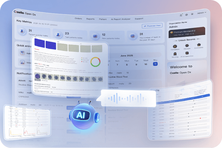
Beyond raw counts and indices, modern veterinary analyzers increasingly incorporate AI-assisted interpretation layers.
By analyzing patterns across multiple parameters, AI models can support the classification of inflammatory profiles and suggest potential directions for further clinical evaluation, such as bacterial versus viral infection patterns or immune-mediated processes.
Result reports may include structured notes explaining how specific deviations in neutrophils, lymphocytes, monocytes, or acute-phase proteins relate to common veterinary pathophysiological mechanisms, while emphasizing that final decisions must be made by clinicians.
These analyzers are also designed to integrate with cloud-based or local IoT platforms that connect devices across multiple veterinary sites.
Connectivity features such as LIS interfaces, LAN ports, and cloud dashboards enable centralized management of instruments, remote monitoring of status, and consolidated quality control.
For veterinary groups operating networks of clinics and laboratories, the automated animal hematology analyzer becomes a node in a larger digital infrastructure for diagnostics and data governance.
Veterinary AI diagnostics providers present these integrated capabilities through an AI-enabled veterinary diagnostics platform that outlines their approach to AI and IoT in animal health.
From this portal, users can access detailed information about veterinary hematology and multi-functional analyzers and understand how device-level AI connects with cloud-level management tools.
Application scenarios in veterinary practice
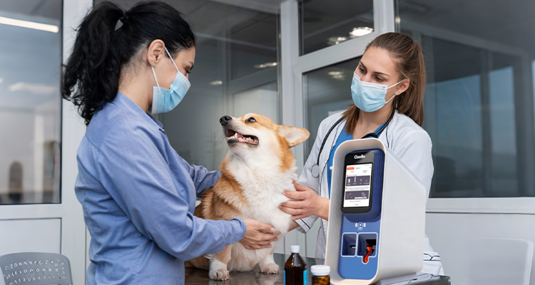
In primary veterinary clinics, automated analyzers support same-visit decision-making by delivering CBC and related results within minutes, allowing clinicians to discuss findings with pet owners during the consultation.
The combination of automated sample handling and AI-based morphology reduces dependence on external laboratories for many routine cases.
For veterinary specialty hospitals and reference laboratories, multi-parameter systems provide an integrated workflow for hematology, urine, fecal, and immunoassay testing on canine and feline samples.
Deployment models can be adapted to the structure of the veterinary organization.
Smaller animal hospitals may focus on CBC and selected immunoassay markers, while regional centers use fully configured analyzers such as the EHVT-50 automated veterinary CBC platform for extended panels, urine sediment, and fecal parasite evaluation.
Through IoT platforms, results from multiple analyzers can be aggregated, compared, and audited centrally, supporting standardized practice and continual improvement of AI algorithms in real-world veterinary workflows.
Technology profile of a modern veterinary automated animal hematology analyzer
| Aspetto | Modern veterinary analyzer (e.g., EHVT-50) |
| Core methodology | AI cell morphology for blood, urine, and feces, plus immunofluorescence for immunoassay. |
| Species focus | Canine and feline, with options for additional species expansion. |
| Sample types | Whole blood, urine, feces, plus immunoassay on veterinary specimens. |
| Differential capability | 7-part CBC with extended morphological flags for abnormal cells. |
| Cartridges and reagents | Single-use wet-staining test kits and dry-type QC cards. |
| Maintenance concept | Maintenance-free operation with sealed reagents and internal waste handling. |
| Produttività | Approximately 8 samples per hour for in-clinic workflows. |
| Connettività | LAN, USB, LIS, and cloud platform integration for remote management. |
This profile summarizes the veterinary-focused architecture and workflow described in the source materials.
Example of an integrated veterinary platform
Within the veterinary portfolio, the EHVT-50 is positioned as a multi-functional analyzer that combines AI-driven 7-differential hematology with urine, fecal, and immunoassay testing for dogs and cats.
It exemplifies how a single automated animal hematology analyzer can serve as a central diagnostic hub in an animal hospital or veterinary laboratory, covering a wide range of routine and specialized tests.
Veterinary professionals seeking more details on such systems can explore the EHVT-50 veterinary hematology analyzer section from the English-language platform, where the test menu, optical architecture, and integration with quality control workflows are described in more depth.
This information sits within a broader overview of AI-driven veterinary diagnostics that highlights how hematology, urine, fecal, and immunoassay testing are brought together in a single veterinary solution.
Conclusione
The evolution of the automated animal hematology analyzer in veterinary diagnostics reflects a broader movement toward automated, AI-assisted, and connected laboratory systems.
By integrating CBM-based imaging, standardized staining, and deep learning algorithms with single-use cartridges and IoT connectivity, modern veterinary analyzers provide both morphological depth and operational simplicity for canine and feline care.
As these systems continue to expand their test menus and integrate with cloud platforms, they are likely to play an increasingly central role in standardizing veterinary hematology, improving data quality across animal hospitals and laboratories, and enabling more data-driven decision-making in everyday practice.