
 English
 Français
 Русский
 Español
 Deutsch
 Português
 العربية
 Italiano

A complete blood count remains one of the most widely used laboratory tests because it provides a rapid overview of major blood-cell populations and core parameters. However, as laboratory workloads grow and clinical teams require faster, more interpretable information, numerical outputs alone do not resolve every review question—particularly when abnormal flags or atypical cell populations require closer examination.
AI-assisted digital morphology is becoming a more visible part of modern hematology workflows, although implementation architectures vary. Some systems integrate imaging and classification directly with CBC analysis, while others connect automated hematology with separate digital morphology platforms. Across these architectures, AI can support cell recognition, classification, and organization of morphology-related information. In integrated image-based hematology systems, CBC measurement, cell imaging, AI-assisted classification, and image-backed review may be brought together within the same analytical workflow.
For hospitals and laboratories evaluating this shift, the question is broader than whether an analyzer can generate more parameters. It is whether the system can turn blood-cell information into a reviewable, traceable, and operationally useful workflow.
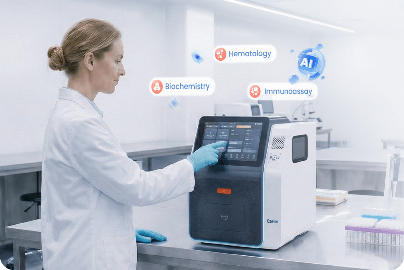
Why CBC Workflows Still Require Morphology Review
Routine CBC Results Do Not Resolve Every Review Question
Modern automated hematology analyzers provide quantitative CBC parameters, leukocyte differentials, flags, histograms, scattergrams, and other system-specific outputs at scale. White blood cell counts, red blood cell indices, hemoglobin, platelet measurements, and differential results remain central to day-to-day patient assessment. These outputs are efficient for high-volume work and provide an important quantitative foundation for hematologic assessment.
However, a numerical result or instrument flag does not always explain the cellular pattern behind it. Samples with unusual scatter characteristics, abnormal differential patterns, immature cells, atypical lymphocytes, or morphology-related concerns may require further review. In a traditional laboratory pathway, that escalation often leads to peripheral blood smear preparation and manual microscopic examination.
Morphology Review Depends on Specialist Time
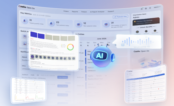
Manual morphology review retains substantial clinical value, but it is resource-intensive. Accurate identification of cell type, maturity, nuclear characteristics, cytoplasmic features, and atypical patterns depends on training and repeated experience. Review workload also varies significantly: a laboratory may process many routine samples efficiently, then face a group of flagged or complex samples requiring concentrated specialist attention.
The challenges of manual morphology analysis extend beyond the technical demands of using a microscope. Laboratories need to maintain a consistent approach to morphology-related review across different shifts, locations, and changing sample volumes. This makes it necessary to focus specialist attention on samples that require closer assessment, while retaining professional control over final interpretation.
Distributed Testing Raises New Workflow Requirements
Hematology testing is no longer limited to large central laboratories. Hospitals, outpatient departments, emergency settings, health-check centers, and distributed care networks all need timely and reliable blood-analysis workflows. These environments differ in sample volume, staffing, physical space, maintenance capacity, and access to morphology expertise.
This shift increases the value of systems that can combine routine CBC measurement with clear escalation paths for samples that need additional review. For laboratories exploring this transition, human hematology analyzer solutions increasingly reflect a wider industry move toward integrating routine blood analysis with morphology-oriented information rather than treating them as disconnected workflows.
From Numerical Parameters to Digital Cell Information
One important direction in hematology is the addition of digitized cellular morphology to established CBC workflows. Standalone conventional analyzers primarily report quantitative values, percentages, histograms, scattergrams, and flags. Digital morphology systems add visual cell information that can be stored, reviewed, compared, and linked to the numerical result.
That distinction matters because morphology is inherently visual. Cell size, granularity, nuclear shape, chromatin pattern, cytoplasmic appearance, and developmental stage cannot be fully conveyed through a single numerical index. A structured imaging workflow can give laboratory personnel another way to investigate why a result has been flagged or why a sample merits additional attention.
A useful way to understand this evolution is to view it as a layered model:
| Information layer | Primary output | Operational role |
| Routine CBC | Counts, indices, differential values | Establishes the quantitative blood profile |
| Instrument flags and distributions | Alerts, histograms, scatter patterns | Identifies samples that may need further attention |
| Digital morphology | Cell images and morphology-related findings | Adds visual context for review |
| AI-assisted analysis | Preclassification and abnormality prompts | Helps prioritize review and organize evidence |
| Professional review | Reclassification, result review, and authorization | Maintains expert oversight and accountability |
Different architectures handle this layer differently. In downstream digital morphology workflows, analyzer flags or laboratory review rules may determine which smears proceed to digital imaging. In integrated image-based hematology systems, cell imaging and AI-assisted classification can be part of the primary analytical workflow, while laboratory rules determine which results require additional professional review.
What Changes When AI-Assisted Morphology Is Integrated With CBC Testing?

An AI CBC analyzer is not simply a conventional CBC instrument with an AI feature added to it. It represents a workflow approach that brings together cell imaging, algorithmic analysis, result presentation,
and professional review. Its usefulness depends on whether these elements support a clear, reviewable, and practical laboratory process.
Standardized Imaging Is the Foundation
AI-assisted morphology depends on image quality and consistency. If cell images are poorly separated, inconsistently acquired, or difficult to compare, algorithmic classification becomes less useful and professional review becomes slower. Consistent sample preparation, staining, image acquisition, focus, segmentation, and image presentation all affect the quality of digital morphology analysis.
For laboratories, this means that the evaluation of an AI-enabled system should include more than its analytical principle. Review teams should ask how the system presents images, whether cells can be inspected efficiently, how images correspond to reported findings, and how the workflow handles samples that need escalation.
AI Supports Triage, Not Independent Diagnosis
AI can assist with cell detection, image segmentation, preclassification, and the identification of patterns that may warrant review. These capabilities can reduce repetitive visual sorting and help a laboratory concentrate attention where it is most needed.
An ICSH review of digital morphology analyzers describes AI-based cell preclassification as a way to automate parts of blood-smear review and support faster slide assessment. The same review emphasizes that skilled morphologists remain essential for cell reclassification and diagnostic interpretation.
These recommendations primarily address digital blood-smear morphology workflows. Integrated image-based CBC systems may use different analytical architectures, so their morphology outputs and review pathways should be evaluated according to the system’s validated intended use and laboratory procedures.
The practical use case is therefore closer to triage than autonomous diagnosis. A system may organize cells into preliminary groups, highlight images associated with atypical findings, or make it easier to compare morphology-related information with CBC data. A qualified reviewer must still assess the relevance of the finding, assess the finding alongside other laboratory results and relevant clinical information.
This distinction is especially important for abnormal and uncommon samples. Algorithms can support consistency and speed, but local validation, quality controls, review procedures, and human accountability remain necessary components of clinical laboratory practice.
Image-Backed Results Improve Reviewability
A result becomes more useful when the reviewer can see the evidence associated with it. In a morphology-oriented workflow, that may include relevant cell images alongside quantitative findings and AI-assisted categories. Instead of receiving only a generic alert, the laboratory can review a more structured set of information.
This approach can support several practical tasks:
- Reviewing the visual basis for an abnormality prompt
- Comparing morphology-related findings with CBC values and differential patterns
- Recording or revisiting images for quality review and staff training
- Supporting communication between laboratories or between laboratory and clinical teams when an image-based discussion is needed
Digital morphology also has broader operational uses. Image archiving, remote review, competency assessment, quality assurance, and educational workflows can all benefit from a system that converts transient microscope observations into organized digital information.
The Workflow Impact: Where AI Adds Value
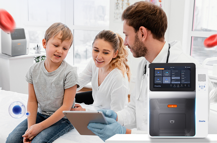
Laboratory workflows vary by sample mix, review criteria, staffing, and information-system design. AI-assisted morphology can therefore enter the CBC workflow through different architectures. Two broad models help illustrate the distinction: downstream digital morphology after CBC screening and integrated image-based morphology within the primary hematology workflow.
Model 1: Rule-Triggered Digital Morphology
In a downstream digital morphology workflow, routine CBC analysis is performed first. Analyzer flags, abnormal distributions, or laboratory-defined review criteria determine which samples require additional morphology assessment.
A typical sequence may include:
- Sample receipt and identification: The specimen enters the laboratory through established accessioning and identification procedures.
- Routine CBC analysis: The analyzer generates quantitative results, differential information, graphical outputs, and instrument flags.
- Review-rule selection: Laboratory-defined criteria determine whether the sample requires additional morphology review.
- Smear preparation and digital imaging: Selected samples proceed to blood-smear preparation, imaging, and AI-assisted cell preclassification where applicable.
- Professional morphology review: Qualified laboratory personnel review and, where necessary, reclassify cells or request additional microscopy or testing.
- Result review and reporting: Findings are reviewed according to laboratory procedures before the result is authorized and released.
In this model, digital morphology is introduced after the initial CBC result, primarily for samples that meet defined review criteria.
Model 2: Integrated Image-Based Hematology
In an integrated image-based hematology workflow, cell imaging and AI-assisted classification are incorporated into the primary analytical process rather than added only after a CBC flag.
A typical sequence may include:
- Sample receipt and identification: The specimen enters the established laboratory workflow.
- CBC analysis and cell imaging: Quantitative hematology results and cell-image information are generated within the same analytical workflow.
- AI-assisted classification: Algorithms organize or classify image-based cellular information according to the system’s validated capabilities.
- Laboratory-defined review: Quantitative results, flags, morphology-related findings, and corresponding images are reviewed together. Laboratory rules determine which results require additional professional attention.
- Additional review where required: Manual smear review, microscopy, repeat testing, or other follow-up procedures may still be used when automated findings are uncertain, discordant with other laboratory information, or meet established review criteria.
- Result review and reporting: Qualified personnel apply laboratory procedures before results are authorized and released.
In this model, image-based morphology is already available within the initial hematology workflow. The laboratory does not necessarily decide whether images are generated after a flag; instead, its review rules determine which results require additional professional assessment.
The Difference Is Where Morphology Enters the Workflow
Both approaches retain professional oversight. The key distinction is the point at which morphology-related information becomes available.
| Workflow architecture | When morphology enters | Role of laboratory review rules |
| Downstream digital morphology | After CBC screening, for selected samples | Determines which samples proceed to smear imaging and morphology review |
| Integrated image-based hematology | During the primary CBC analytical workflow | Determines which image-backed results require additional professional review or follow-up |
AI therefore does not replace laboratory review in either model. Its role is to help organize, classify, and present morphology-related information within the architecture used by the laboratory. The practical value depends on whether that information is integrated into a clear review process, supported by appropriate quality procedures, and interpreted by qualified personnel.
Application Scenarios for AI-Assisted CBC Workflows
Different healthcare settings face different constraints, so the value of an AI CBC analyzer will vary by use case. The same technology should not be assessed with a single “more automation is better” standard.

Central Laboratories
High-volume central laboratories may benefit most from structured exception handling. The priority is often not merely producing results quickly, but ensuring that flagged and complex samples move efficiently to appropriate review. AI-assisted preclassification and image-backed findings can help reviewers focus on samples with greater morphology-related relevance.
For these laboratories, integration matters. A morphology platform that sits outside the main result-review process may add another manual handoff. The operational value increases when quantitative data, images, review tools, and laboratory information-system workflows are coordinated.
Hospital Departments and Time-Sensitive Testing
Emergency departments, inpatient services, hematology units, and oncology-related care settings may place particular value on timely recognition of samples that warrant escalation. The analyzer does not make a diagnosis, but it can help the laboratory identify which results should be reviewed with greater urgency.
The relevant requirement is clarity. Reviewers need a result format that brings together cell counts, relevant flags, morphology-related prompts, and accessible images without forcing them to navigate several disconnected interfaces. A clear workflow can support faster review decisions while maintaining appropriate professional checks.
Distributed and Resource-Constrained Settings
Smaller laboratories, outpatient facilities, and distributed healthcare networks may have limited access to staff with extensive morphology experience. In these settings, compact automation and standardized imaging can help make morphology-related information more accessible within a routine testing process.
Digital images may also create a foundation for remote consultation or centralized review, where laboratory governance and local procedures allow it. This does not remove the need for trained professionals. It can, however, make specialist expertise easier to direct toward the samples and sites that need it most.
From Industry Trend to Deployable System
The move toward morphology intelligence is influencing how hematology platforms are designed. In addition to routine analysis, laboratories increasingly expect image-backed review, AI-assisted recognition, automation, and information connectivity to work together rather than exist as separate components.
Ozelle applies this approach through its AI × Complete Blood Morphology (AI × CBM) framework, which brings AI-assisted morphology and cell-image evidence into human hematology workflows. The framework reflects the wider move from isolated parameter reporting toward more structured, reviewable morphology information.
Within this framework, the O-Cyte 1 automated hematology analyzer combines 7-diff CBC testing with AI-assisted morphology in a single workflow. It automatically recognizes and classifies blood cells, linking quantitative findings and morphology-related results to corresponding cell images. This format gives laboratory professionals a more direct route from an abnormality prompt to image-backed evidence for review.
For laboratories considering how morphology intelligence fits their own testing environment, Ozelle’s hematology solutions provide a reference point for examining how routine CBC analysis, image-based information, and AI-assisted review can be combined in a single operational model.
The Next Phase of CBC Analysis
The next phase of hematology is likely to depend not on AI alone, but on how effectively quantitative CBC analysis, morphology, digital evidence, and professional review can be integrated.
An AI-assisted image-based hematology workflow can add visual morphology evidence to the quantitative, differential, graphical, and flag-based information already produced by automated CBC systems. The opportunity is not to remove professional expertise from the process, but to make morphology information more accessible, reviewable, and better integrated into laboratory-defined procedures.